Microscopy
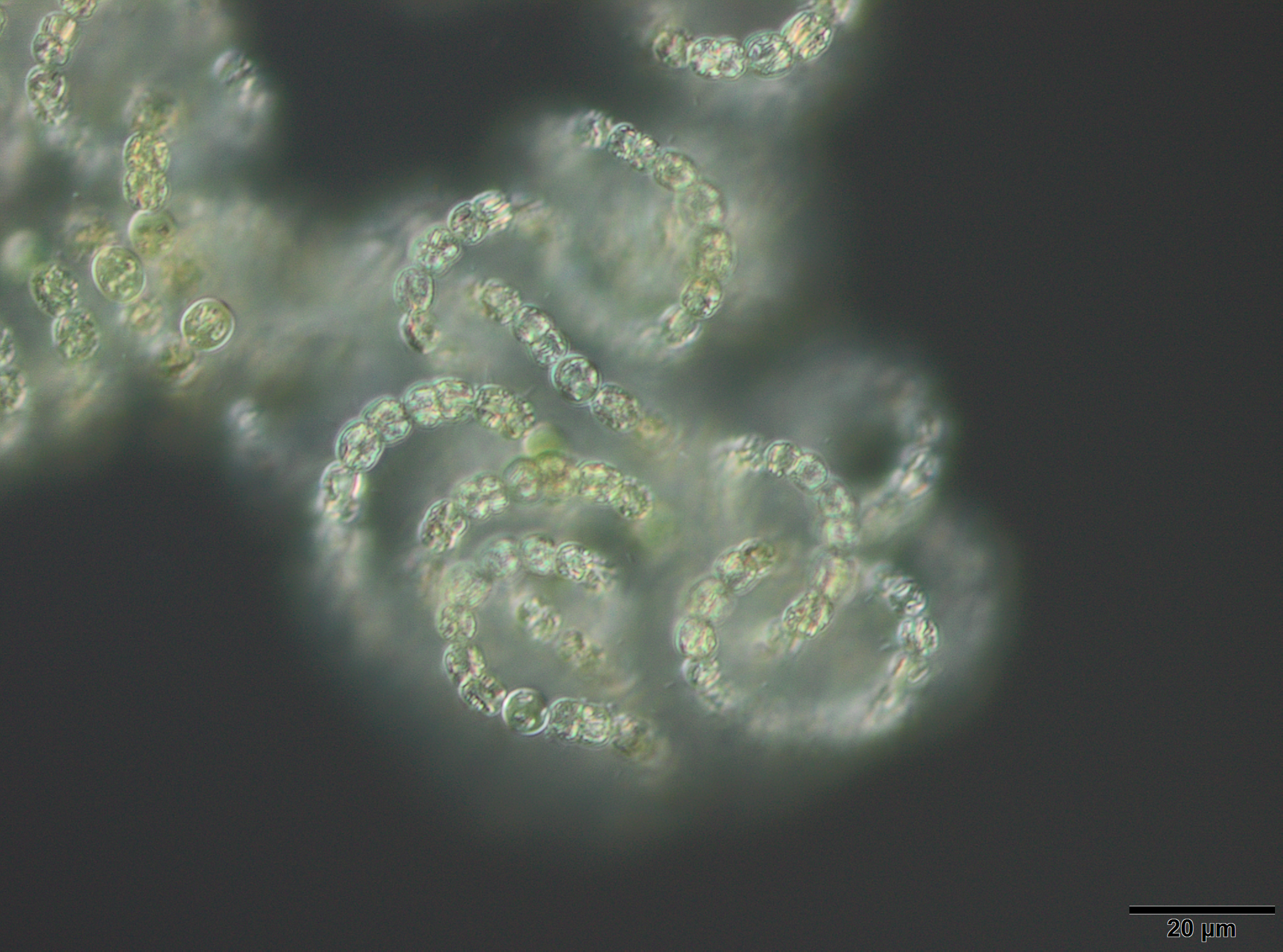
Light Microscopy
The microscopy laboratory is not only used by the Applied Ecology & Phycology but also by the Ecology group and the Cell Biology of Phototrophic Marine Organisms group. Jun. Prof. Dr. Klaus Herburger (AG Cell Biology of Phototrophic Marine Organisms) is an expert in cell biological studies and many different microscopic techniques (e.g. electron microscopy, confocal laser scanning microscopy...). The two Olympus microscopes BX-51 and IX-70 are equipped with differential interference contrast, which allows the visualization of very small cells and cell compartments. The maximum magnification is 1000 times. The inverted microscope IX-70 furthermore provides the possibility to investigate surfaces of bigger objects (e.g. biofilms on soil crusts). Micrographs can be taken, analyzed and saved with an imaging software package. The light microscopy is especially used for morphological determination of living algae and permanent slides of diatom frustules.
BORCHHARDT, N., SCHIEFELBEIN, U., ABARCA, N., BOY, J., MIKHAILYUK, T., SIPMAN, H. & KARSTEN, U. (2017) Diversity of algae and lichens in biological soil crusts of Ardley and King George Island, Antarctica. Antarctic Science 29, 229-237. DOI: 10.1017/S0954102016000638
ALBRECHT, M., PRÖSCHOLD, T. & SCHUMANN, R. (2017) Identification of cyanobacteria in a eutrophic coastal lagoon on the southern Baltic coast. Frontiers in Microbiology 8, 923. DOI: 10.3389/fmicb.2017.00923
SCHULZ, K., MIKHAILYUK, T., DRESSLER, M., LEINWEBER, P. & KARSTEN, U. (2016) Biological soil crusts from coastal dunes at the Baltic Sea: cyanobacterial and algal biodiversity and related soil properties. Microbial Ecology 71, 178-193. DOI: 10.1007/s00248-015-0691-7
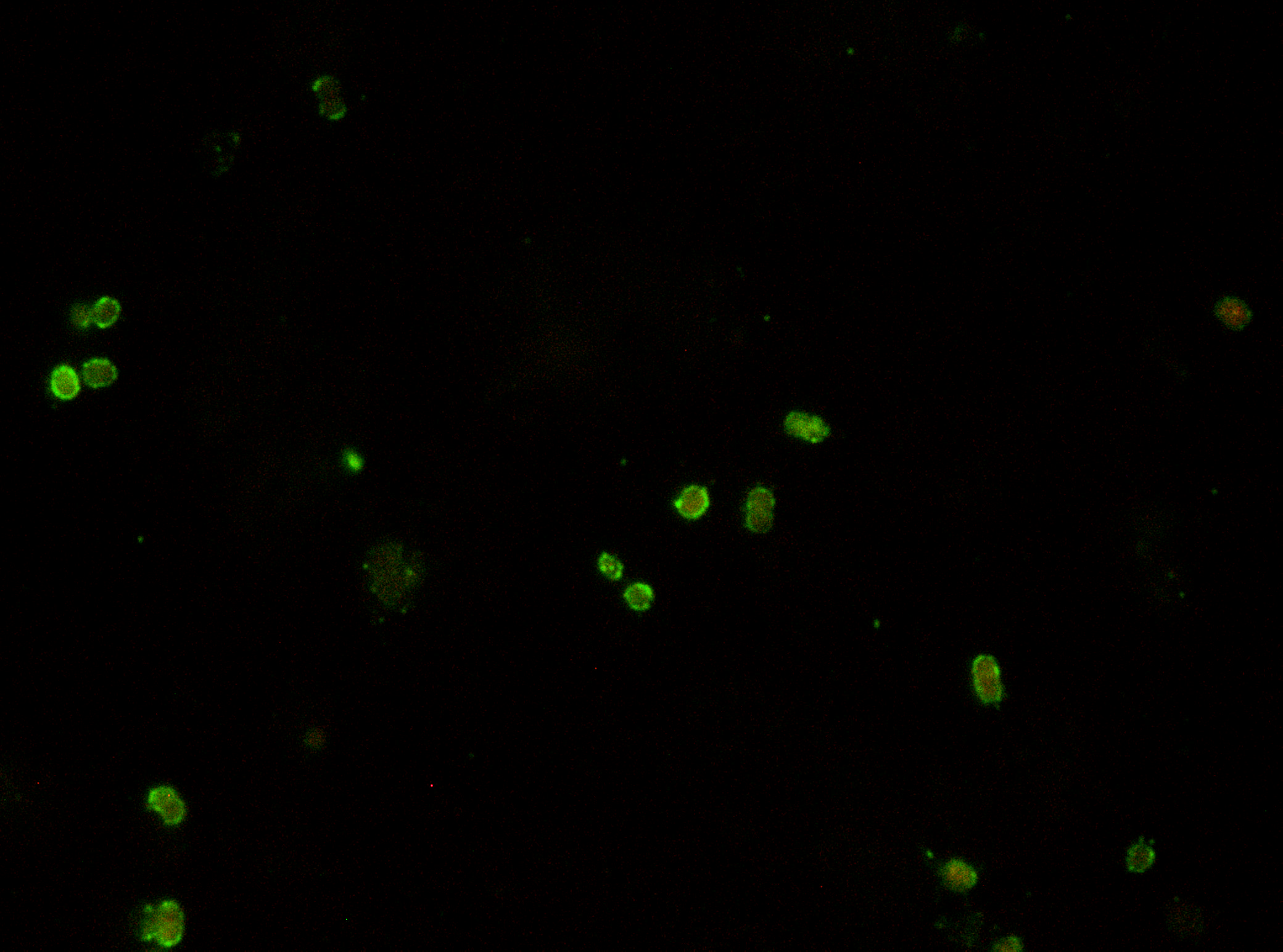
Epi-fluorescence Microscopy
Both light microscopes BX-51 and IX-70 are equipped with epifluorescence, which provide excitation at green, blue and UV spectra. Therefore, a broad range of fluorophores can be used. DAPI-, FITC- and autofluorescence are most frequently applied.
GLADIS, F. & SCHUMANN, R. (2011) A suggested standardised method for testing photocatalytic inactivation of aeroterrestrial algal growth on TiO2-coated glass. International Biodeterioration & Biodegradation 65, 415-422. DOI: 10.1016/j.ibiod.2011.01.005
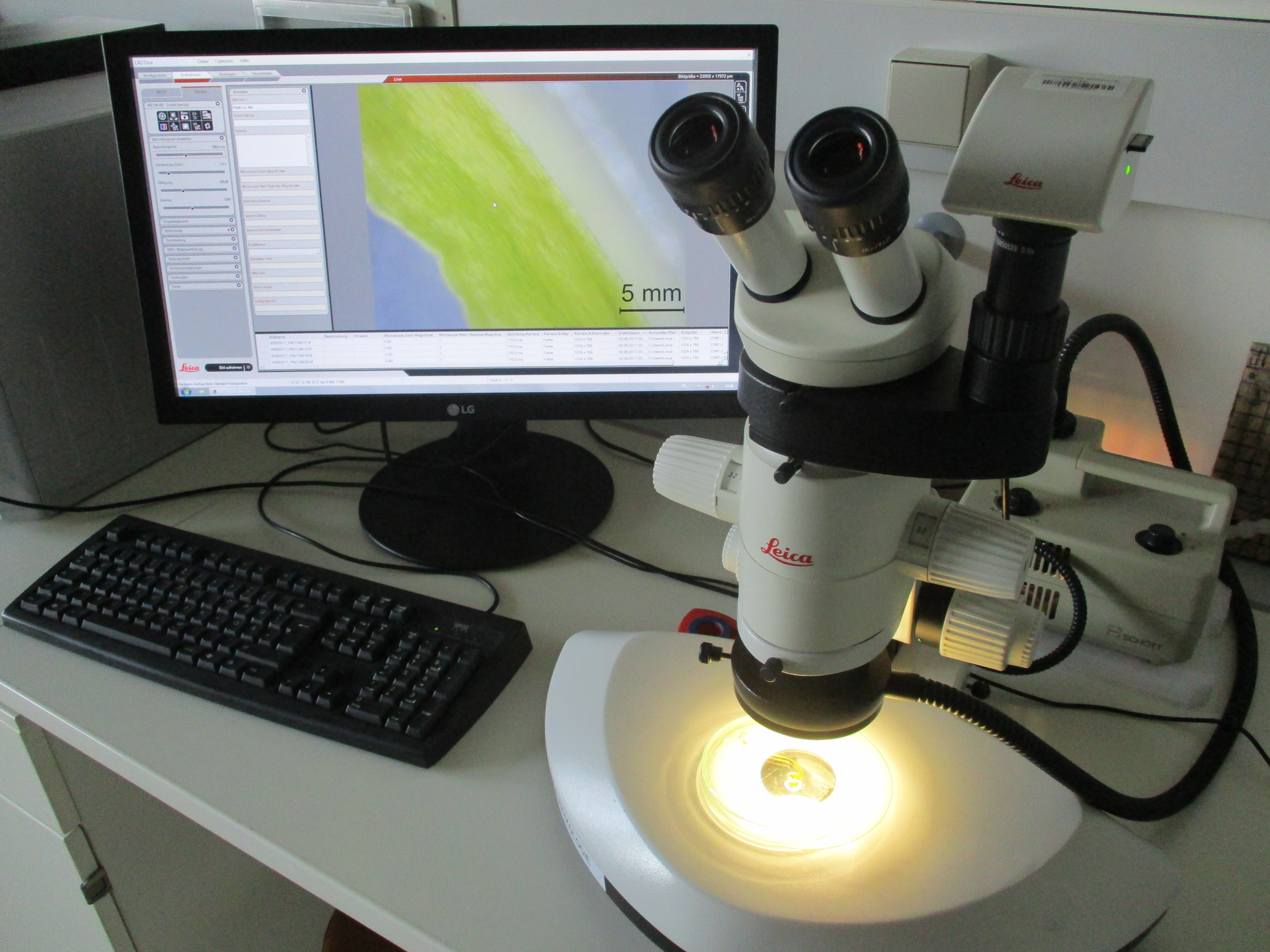
Stereomicroscope
The working group furthermore has a Leica stereomicroscope with a maximum magnification of 64 times to screen larger objects as shipworm tunnels. This stereomicroscope is also connected to an imaging software.
WEIGELT, R., LIPPERT, H., BORGES, L., APPELQVIST, C., KARSTEN, U. & BASTROP, R. (2016) First time DNA barcoding of the common shipworm Teredo navalis Linnaeus 1758 (Mollusca: Bivalvia: Teredinidae): molecular-taxonomic investigation and identification of a widespread wood-borer. Journal of Experimental Marine Biology and Ecology 475, 154-162. DOI: 10.1016/j.jembe.2015.11.008
A number of student microscopes and stereomicroscopes are provided in the laboratories of the working group.